
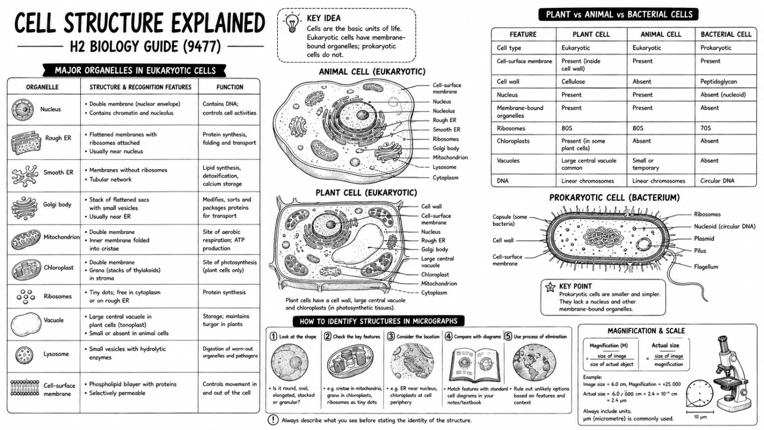
Quick answer: Eukaryotic cells (plant and animal) contain membrane-bound organelles that separate incompatible reactions and create specialised internal conditions; prokaryotic (bacterial) cells don’t. Plant cells have a cellulose cell wall, often a large central vacuole, and chloroplasts in photosynthetic tissue — but they still have a cell-surface membrane inside the wall, controlling exchange the same way an animal cell’s membrane does. This guide covers every required organelle, how to identify structures in real micrographs (not just idealised diagrams), and the comparisons examiners test most often.
Cell structure is easier to learn when every organelle is treated as part of a working system rather than a label to memorise. A diagram may help you remember where a structure sits, but exam questions usually ask how its features support a particular function — which is the reasoning this guide is built around. It follows the Singapore-Cambridge H2 Biology syllabus 9477 for the first examination in 2026 and pairs with the broader H2 Biology Syllabus guide if you need the full Core Idea map that this topic sits within.
The most useful revision pattern is simple: identify the structure, describe the feature that matters, and connect that feature to the biological process it supports.
What Is Cell Structure?
Cell structure describes the organisation of a cell and the specialised parts that keep it alive. Some structures form boundaries, some store genetic information, and others create the conditions needed for chemical reactions.
Eukaryotic cells contain membrane-bound organelles — internal compartments that separate incompatible reactions, concentrate enzymes, and create different conditions within the same cell. Animal and plant cells are both eukaryotic, sharing a nucleus, mitochondria, endoplasmic reticulum, Golgi body, and ribosomes.
Exam focus: don’t stop at naming an organelle. A complete answer usually needs a structure-function link — folded inner membranes providing a large surface area for proteins involved in oxidative phosphorylation, for example, not just “mitochondria make energy.”
The Cell Theory
Cells are the smallest units of life; all cells arise from pre-existing cells; living organisms are composed of cells. Viruses challenge simple definitions because they contain genetic material and can evolve, but they aren’t cellular and can’t reproduce independently — they depend on a host cell for replication, protein synthesis, and many metabolic processes.
Animal and Plant Cell Structure
Animal cells have flexible outer boundaries, since the cell-surface membrane isn’t surrounded by a rigid wall — their shape varies with function, so a neurone, muscle cell, and secretory cell can look quite different despite sharing the same basic organelles.
Plant cells share the eukaryotic organelles found in animal cells, but add a cellulose cell wall. Many mature plant cells have a large central vacuole, while photosynthetic tissues contain chloroplasts. The cell wall sits outside the cell-surface membrane, so plant cells still use the membrane to control movement into and out of the cytoplasm — the wall doesn’t replace that function.
| Feature | Plant cell | Animal cell |
| Cell-surface membrane | Present inside the cell wall | Present as the outer cell boundary |
| Cellulose cell wall | Present | Absent |
| Nucleus | Present in most living cells | Present in most living cells |
| Chloroplasts | Present in photosynthetic tissues | Absent |
| Large central vacuole | Common in mature cells | Absent as a large permanent structure |
| Mitochondria | Present | Present |
| Centrioles | Usually not shown in a typical plant-cell diagram | Present in the syllabus: animal-cell model |
| Shape | Often more regular because of the wall | Often flexible and variable |
Common mistake: plant cells don’t replace the cell-surface membrane with a cell wall — they have both structures, and each has a different function.
Cell Organelles and Their Functions
The organelle list is easier to remember grouped by task: the nucleus stores information, the endomembrane system produces and transports materials, energy-transforming organelles provide usable energy, and the cell boundary manages exchange.
Nucleus, nuclear envelope, and nucleolus: the nucleus holds most of the cell’s DNA as chromatin. The nuclear envelope is a double membrane with pores regulating exchange between the nucleus and cytoplasm, allowing transcription and RNA processing to occur in a controlled compartment. The nucleolus produces ribosomal RNA and assembles ribosomal subunits — often visible as a darker region in a micrograph.
Ribosomes: the sites of translation. Free ribosomes produce proteins that usually function in the cytosol; ribosomes attached to the rough endoplasmic reticulum produce proteins entering the endomembrane system. Eukaryotic cytoplasmic ribosomes are 80S; bacterial ribosomes are 70S — and ribosomes aren’t surrounded by a membrane, so calling them “membrane-bound organelles” is incorrect.
Rough and smooth endoplasmic reticulum: rough ER is flattened membrane sacs with attached ribosomes, important for synthesising, folding, and initially modifying proteins destined for secretion, membrane insertion, or specific organelles. Smooth ER lacks attached ribosomes, and its functions include lipid synthesis and, in some cells, detoxification or calcium storage.
Golgi body and vesicles: stacks of flattened membrane sacs that receive material from the ER, modify it, sort it, and package it into vesicles. A secreted protein follows a linked pathway — rough ER ribosome → transport vesicle → Golgi body → secretory vesicle → fusion with the cell-surface membrane. Learn the order and each stage’s role, not just the organelle names.
Mitochondria: the main sites of aerobic respiration. The inner membrane folds into cristae, providing a large surface area for electron-transfer proteins and ATP synthase; the matrix holds enzymes for the link reaction and Krebs cycle. In an electron micrograph, look for a double boundary and internal folds, not just an oval outline.
Chloroplasts carry out photosynthesis. Thylakoid membranes hold photosynthetic pigments and light-dependent reaction components; stacked thylakoids form grana, while the stroma holds Calvin-cycle enzymes. A chloroplast isn’t simply “a green mitochondrion” — both transform energy and have extensive internal membranes, but with different structures, reactants, and products.
Lysosomes: membrane-bound vesicles containing hydrolytic enzymes, digesting materials taken into the cell, breaking down damaged components, and keeping potentially harmful enzymes separated from the cytoplasm.
Centrioles: cylindrical structures built from microtubules, helping organise microtubules and associated with spindle formation during animal-cell division — often shown as a pair positioned at an angle in diagrams.
Cell-surface membrane and cytoplasm: the membrane forms a selectively permeable boundary, controlling exchange, holding receptors, and supporting cell recognition. The cytoplasm — cytosol plus suspended structures — provides the environment for many enzyme-controlled reactions.
Cellulose cell wall and vacuole: the wall’s cellulose microfibrils provide tensile strength, supporting the cell, maintaining shape, and resisting excessive expansion when water enters. A large central vacuole contains cell sap and contributes to turgor, pushing the cytoplasm toward the cell boundary — don’t confuse it with an empty-looking space caused by poor staining.
Quick Organelle Comparison Table
| Structure | Recognition feature | Main function |
| Nucleus | Large compartment with a double envelope; may show a darker nucleolus | Stores DNA, controls gene expression |
| Rough ER | Flattened membranes with small ribosome dots | Produces and processes proteins entering the endomembrane system |
| Smooth ER | Tubular membranes without ribosome dots | Synthesises lipids, supports specialised metabolism |
| Golgi body | Stacked curved sacs with nearby vesicles | Modifies, sorts, and packages materials |
| Mitochondrion | Double boundary with folded inner membrane | Aerobic respiration and ATP production |
| Chloroplast | Double boundary with thylakoid stacks | Photosynthesis |
| Lysosome | Small membrane-bound vesicle, dense contents | Digests material using hydrolytic enzymes |
| Ribosome | Very small particle; free or attached to rough ER | Translates mRNA into a polypeptide |
| Centriole | Paired cylinders made from microtubules | Helps organise spindle microtubules |
Bacterial Cell Structure
Bacterial cells are prokaryotic: small, unicellular, with a peptidoglycan cell wall, circular DNA, and 70S ribosomes. The DNA sits in the cytoplasm rather than inside a nucleus. The absence of membrane-bound organelles doesn’t mean the cell lacks organisation — its cell-surface membrane manages exchange while the cytoplasm holds the enzymes and ribosomes needed for metabolism and protein synthesis.
| Feature | Plant cell | Animal cell | Bacterial cell |
| Cell type | Eukaryotic | Eukaryotic | Prokaryotic |
| Nucleus | Present | Present | Absent |
| DNA | Linear chromosomes in nucleus | Linear chromosomes in nucleus | Circular DNA in cytoplasm |
| Ribosomes | 80S in cytoplasm | 80S in cytoplasm | 70S |
| Membrane-bound organelles | Present | Present | Absent |
| Cell wall | Cellulose | Absent | Peptidoglycan |
| Mitochondria | Present | Present | Absent |
| Chloroplasts | Present in photosynthetic tissue | Absent | Absent |
How to Identify Organelles in Micrographs
Real micrographs don’t look as neat as textbook diagrams — structures may be cut at an angle, partly hidden, or shown at an unfamiliar scale. Base identification on visible evidence:
- Start with the boundary. A thick outer wall suggests a plant or bacterial cell; a flexible membrane alone is consistent with an animal cell.
- Look for internal compartments. A nucleus and other membrane-bound organelles indicate a eukaryotic cell.
- Use internal detail. Cristae support a mitochondrion identification; grana support a chloroplast identification.
- Check for attached ribosome dots. Rough ER has them; Golgi stacks don’t.
- Use scale before deciding. A small dot at low magnification may not contain enough detail for a confident organelle label.
Exam wording: write “identified as a mitochondrion because folded inner membranes or cristae are visible,” not just “it is oval.” Shape alone is weak evidence.
Magnification and Cell Size
Cell-structure questions may combine recognition with measurement. Image size and actual size must use the same unit before calculating.
Magnification = image size ÷ actual size. Actual size = image size ÷ magnification
Convert millimetres to micrometres (× 1,000), micrometres to nanometres (× 1,000). Show working, include units, and use an appropriate number of significant figures. An eyepiece graticule has no fixed unit until calibrated with a stage micrometer at the selected objective lens — changing the objective changes the calibration.
Common Cell-Structure Exam Mistakes
| Mistake | Why it loses marks | Better approach |
| Listing an organelle without a function | The question may need explanation, not just identification | Link one visible feature to the process it supports |
| Calling ribosomes membrane-bound | Ribosomes have no surrounding membrane | Describe them as particles made from rRNA and proteins |
| Saying bacterial cells have no DNA | They lack a nucleus, not genetic material | State that circular DNA lies in the cytoplasm |
| Saying only plant cells have membranes | Plant cells have a membrane inside the wall | Separate the selectively permeable membrane from the supporting wall |
| Identifying mitochondria by shape alone | Many components can look oval in section | Use cristae or a double boundary as evidence |
| Assuming every plant cell has chloroplasts | Non-photosynthetic tissues may not contain them | Refer to photosynthetic plant cells when chloroplasts are relevant |
| Confusing Golgi body with rough ER | Both contain flattened membranes | Check for attached ribosomes and nearby budding vesicles |
| Ignoring units in magnification | Different units create an incorrect value | Convert first, calculate second, state the final unit |
A Step-by-Step Exam Method
- Read the command word and decide whether the task is identify, describe, compare, or explain.
- Use the diagram or micrograph as evidence instead of recalling an idealised picture.
- Name the structure precisely — inner membrane, nuclear envelope, peptidoglycan — where relevant.
- Connect the feature to a biological process with a clear cause-and-effect sentence.
- Check units, labels, and comparison language before moving on.
For a comparison, keep the same feature in both halves of the sentence: “A plant cell has a cellulose cell wall, while an animal cell does not” is clearer than two unrelated lists.
Cell Structure Revision Checklist
□ I can outline the three statements in the cell theory. I can label a typical animal cell and plant cell without notes. I can explain the function of every required organelle. I can link the nucleus, ribosomes, rough ER, Golgi body, and vesicles in a protein-secretion pathway. I can distinguish mitochondria from chloroplasts using internal membranes. I can compare plant, animal, and bacterial cells feature by feature. I can explain why a bacterial cell has DNA but no nucleus. I can identify organelles from micrograph evidence rather than shape alone. I can calculate magnification or actual size after converting units. I can explain why viruses challenge the cell theory.
Cell Structure FAQs
1. What are the main cell organelles for H2 Biology?
The 2026 syllabus names rough and smooth ER, Golgi body, mitochondria, ribosomes, lysosomes, chloroplasts, the cell-surface membrane, nuclear envelope, centrioles, nucleus, and nucleolus.
- Grouping them by function (information, transport, energy, boundary) makes them easier to retain than a flat list.
2. What’s the main difference between a plant cell and an animal cell?
A typical plant cell has a cellulose cell wall and may contain chloroplasts or a large central vacuole; an animal cell lacks both.
- Both are eukaryotic and share most membrane-bound organelles — the differences are additions, not a different basic plan.
3. What’s the difference between a prokaryotic and a eukaryotic cell?
A eukaryotic cell has a nucleus and membrane-bound organelles; a prokaryotic bacterial cell has circular DNA in the cytoplasm, 70S ribosomes, and no membrane-bound organelles.
- “No nucleus” doesn’t mean “no DNA” — this is one of the most common exam mix-ups.
4. How do I remember organelle functions?
Group structures into systems rather than memorising each in isolation: an information pathway (nucleus, ribosomes), a protein pathway (rough ER, Golgi), an energy pathway (mitochondria/chloroplast), and boundary functions (membranes, walls).
- This mirrors how exam questions actually test the material — as connected systems, not isolated facts.
5. Do I need to draw perfect cell diagrams?
No — a biological drawing should be clear, proportionate, and labelled with straight lines; accuracy matters more than artistic detail or shading
6. Are viruses cells?
No — viruses are acellular. They contain genetic material and can evolve, but depend on host cells for reproduction and many life processes.
- This is exactly why viruses are used to test understanding of the cell theory itself.
7. Is cell structure tested at O-Level Biology too, and how does H2 go further?
Yes — O-Level introduces the main organelles and plant/animal comparisons, while H2 adds micrograph identification from real evidence, prokaryote/eukaryote precision, and the expectation that structure links directly to function in every answer.
- A student who only memorised organelle names at O-Level without the structure-function habit often struggles when H2 questions ask for the reasoning, not just the label.
- Our O-Level Biology Tips to Score A1 guide covers building that habit at O-Level depth.
8. Is naming an organelle without explaining its function a common mistake?
Yes — it’s one of the most frequent ways marks are lost here, since “mitochondria make energy” states a fact without the structure-function link (folded inner membranes, large surface area) that full marks usually require.
- Our Common Biology Exam Mistakes guide covers this pattern in more depth, with examples across different Biology topics.
9. How does cell structure connect to enzymes?
An enzyme’s active site is a protein structure, so the same structure-function reasoning covered here — a folded membrane or specific shape enabling a particular job — applies directly to how an active site’s shape determines what it can bind.
- Our Enzymes Explained guide builds on this foundation with the full mechanism, inhibition, and worked rate calculations.
10. Does this apply to O-Level and IP Biology as well as H2?
The core organelles and structure-function reasoning are the same across all three tracks — what changes is depth: H2 adds micrograph identification from real evidence and prokaryote/eukaryote precision that O-Level and early IP Biology don’t require.
- O-Level Biology and IP Biology both introduce cell structure before this level of reasoning is expected.
More Articles from Pamela’s Place
Get Exam-Ready with Pamela’s Place
Cell structure supports later topics — membranes, respiration, photosynthesis, gene expression, cell division. If the organelle links are weak, later explanations feel like separate facts instead of one connected system. At Pamela’s Place, Biology revision is organised around labelled-diagram recall, structure-function explanations, micrograph practice, and targeted correction of common mistakes, in small Omakase groups capped at 7 students — the aim is more precision, not more memorisation.

2 thoughts on “Cell Structure Explained: H2 Biology Guide (9477)”